Our Location
304 North Cardinal St.
Dorchester Center, MA 02124

304 North Cardinal St.
Dorchester Center, MA 02124

Immunohistochemistry (IHC) is a widely used technique that enables visualization of protein expression directly within preserved tissue architecture while maintaining its microstructure. By using specific primary antibodies, IHC allows researchers to determine the localization, distribution, and relative abundance of target proteins within tissue sections, making it highly valuable for studying biological processes in situ, including comparisons between healthy and diseased tissues and understanding molecular changes in their native histological context. Detection is commonly achieved through chromogenic systems such as DAB or fluorescence-based labeling via secondary antibodies, enabling clear visualization of antigen–antibody interactions under a microscope. High-quality, pathology-validated IHC antibodies and workflow solutions are also available from Abclonal, supporting reliable and reproducible immunohistochemistry applications in research and translational studies, with official distribution in Indonesia handled by PT. Kairos Jaya Sejahtera, where products can be ordered directly.
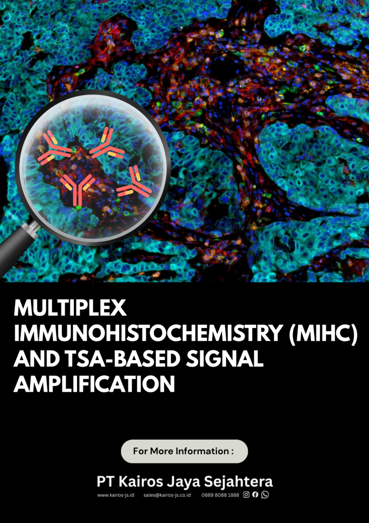
However, conventional IHC is inherently limited in its multiplexing capacity, typically allowing only one to three biomarkers to be analyzed per tissue section. This limitation makes it difficult to fully capture the complexity of biological systems where multiple signaling pathways, diverse cell populations, and dynamic cellular interactions occur simultaneously within the same microenvironment. To address this challenge, Multiplex Immunohistochemistry (mIHC) has emerged as an advanced spatial proteomics approach that enables simultaneous detection of multiple protein targets—commonly ranging from 3 up to 10 or more markers—within a single tissue section. By integrating high-resolution imaging with computational analysis, mIHC provides a multidimensional spatial map of tissue organization, allowing researchers to decode cellular phenotypes, functional states, and intercellular interactions in situ. This approach is based on tyramide signal amplification (TSA) combined with sequential immunostaining cycles, enabling repeated rounds of antibody labeling while preserving tissue integrity and spatial resolution.
In this context, TSA-based multiplex fluorescence systems such as the TSA Fluorescence Triple Staining Plus Kit (Rabbit and Mouse Secondary Antibodies) (Abclonal RK05903P https://abclonal.com/catalog-antibodies/TSAFluorescenceTripleStainingPlusKitRabbitandmousesecondaryantibodies/RK05903P) play a critical role in enabling practical implementation of mIHC workflows. This kit utilizes HRP-conjugated secondary antibodies and tyramide signal amplification chemistry to generate highly stable, covalently bound fluorescent signals at the site of antigen detection. Through sequential staining cycles combined with signal inactivation steps, multiple protein targets can be labeled within a single tissue section without significant cross-reactivity, making true triple staining and higher-order multiplexing feasible.
As biomedical research continues to shift toward systems biology and spatially resolved analysis, the need for multiplex and higher-order staining strategies such as triple staining has become increasingly urgent. Many diseases, including cancer, autoimmune disorders, and chronic inflammatory conditions, are not driven by a single biomarker but by complex networks of interacting cells and signaling pathways. Traditional single-marker or low-plex approaches often fail to capture true tissue heterogeneity, require serial sections that introduce variability, and limit the ability to study spatial cell-to-cell interactions. In contrast, triple staining and multiplex IHC enable simultaneous visualization of multiple biomarkers within a single intact tissue section, improving accuracy in mapping immune infiltration, tumor microenvironment architecture, and functional cellular states while reducing tissue consumption.
When comparing methodologies, mIHC clearly extends beyond the limitations of conventional IHC and immunofluorescence (IF).
Feature | mIHC (Multiplex IHC) | IF (Immunofluorescence) | IHC (Conventional) |
|---|---|---|---|
Number of Targets | 3–10+ markers | 1–3 markers | 1–3 markers |
Signal Stability | High stability, covalently deposited signals | Prone to photobleaching and spectral overlap | Stable chromogenic signal |
Spatial Resolution | High-dimensional spatial mapping | High resolution but limited multiplexing | Moderate spatial information |
Antibody Compatibility | No species restriction due to sequential staining | Requires species-matched antibodies | Requires species-matched antibodies |
Staining Strategy | Sequential staining with TSA amplification | Simultaneous staining | Single staining or serial sections |
Detection System | Tyramide signal amplification (TSA) | Fluorophores | DAB / Fast Red |
Multiplex Capability | Excellent (up to 10+ markers) | Limited beyond 3 markers | Very limited |
Data Output | High-dimensional spatial proteomics | Limited multiplex imaging | Qualitative / semi-quantitative |
Overall, TSA-based multiplex immunofluorescence systems such as the Abclonal triple staining kit represent a key enabling technology that bridges conventional IHC workflows with modern spatial biology. This transition allows researchers to move from single-marker histology toward high-dimensional tissue analysis, providing deeper insight into tissue microenvironments, immune landscapes, and disease mechanisms, and supporting the growing demand for precision medicine and translational research.
WhatsApp us